Living model of the oligodendrocyte: the cell. Zoom between the stages.Morphology after Peters, Palay and Webster, The Fine Structure of the Nervous System (1991); Nave and Werner, Annu Rev Cell Dev Biol 2014; Chong et al., PNAS 2012 (internode counts per cell).
Oligodendro, at your service. Everything known about the oligodendrocyte, and rather more about myelin than is strictly decent. What would you like?
The oligodendrocyte is a glial cell of the central nervous system whose principal function is to produce myelin, the lipid-rich membrane that wraps around axons in concentric layers and enables fast, energy-efficient signal transmission. A single oligodendrocyte extends processes to myelinate up to fifty separate axon internodes, a reach that has no parallel in the peripheral nervous system. Beyond insulation, oligodendrocytes supply lactate and pyruvate to axons through monocarboxylate transporters, providing metabolic support that is required for long-term axon survival. When myelin is lost, as in multiple sclerosis, oligodendrocyte precursor cells can be recruited to rebuild it, though this capacity declines with age and fails entirely in chronic lesions. (Nave and Werner 2014, https://pubmed.ncbi.nlm.nih.gov/25288117/; Funfschilling et al. 2012, https://pubmed.ncbi.nlm.nih.gov/22622581/)
Comprehensive review of CNS and PNS myelination: sheath assembly, axon glia signalling, and the metabolic role of the oligodendrocyte. The modern structural reference.
Disabling oxidative phosphorylation in oligodendrocytes does not cause demyelination; the cells run on aerobic glycolysis and pass the product to the axon.
MCT1 in oligodendrocytes exports lactate to axons. Deleting it causes axon degeneration with the myelin sheath still intact, separating support from insulation.
Mouse. After a paw injury, spinal oligodendrocytes switch from making myelin protein to making lipid, axons suffer, and neuronal amyloid-beta42 builds; blocking it prevented the pain becoming chronic.
Anti-CD20 B-cell depletion reduces relapse rate and new lesions in relapsing MS. Oligodendrocyte protection is indirect, by removing the immune attack.
An antimuscarinic antihistamine repurposed as a differentiation promoter. In this crossover trial it shortened visual evoked potential latency in chronic optic neuropathy, the first remyelination signal from a drug in humans.
S1P receptor modulator approved for secondary progressive MS. S1P5 is expressed on oligodendrocytes, so a direct effect is plausible alongside the immune one.
Clemastine fumarateantihistamine with antimuscarinic action
acts on muscarinic receptors (M1 and M3)
Promotes oligodendrocyte precursor differentiation and remyelination. In the ReBUILD crossover trial the primary endpoint was met: visual evoked potential latency delay reduced by 1.7 ms per eye (95% CI 0.5 to 2.9, p = 0.0048); fatigue was associated, no serious adverse events.
dose: trial dose not stated in the abstract · human, 50 patients with multiple sclerosis · randomised double-blind placebo-controlled crossover trial, 150 days · Green AJ et al. 2017, Lancet · source
Clemastineantihistamine with antimuscarinic action
acts on muscarinic receptors, ERK1/2, Myrf and Olig2
Promotes OPC differentiation through muscarinic receptors and ERK1/2 activation with increased Myrf and Olig2; blocked by ERK inhibition (U0126) and reduced by the muscarinic agonist cevimeline.
dose: not stated in the abstract · rat, spinal cord injury · in vivo pharmacology · Tong LY et al. 2022, Front Pharmacol · source
Screening 1,000 bioactive molecules on micropillar arrays identified a cluster of antimuscarinic compounds that enhance oligodendrocyte differentiation and remyelination.
dose: screen concentrations not stated in the abstract · rodent OPCs in vitro (species not stated in the abstract) · high-throughput micropillar screen · Mei F et al. 2014, Nat Med · source
Benztropineanticholinergic
acts on direct antagonism of M1 and/or M3 muscarinic receptors
Among the most effective compounds in an image-based screen for myelin basic protein expression; decreases clinical severity in EAE alone or with approved immunosuppressants; efficacy from enhanced remyelination, not immune suppression (cuprizone model, T-cell assays).
dose: not stated in the abstract · rat optic nerve OPCs in vitro; mouse EAE and cuprizone models · screen plus in vivo models · Deshmukh VA et al. 2013, Nature · source
Miconazoleantifungal
acts on mitogen-activated protein kinase signalling in OPCs
Increases new oligodendrocytes and enhances remyelination in lysolecithin lesions; reverses disease severity at the peak of EAE; acts directly as a remyelinating drug with no immune effect; enhances human oligodendrocyte generation from human OPCs in vitro.
dose: not stated in the abstract · mouse; human cells in vitro · in vivo lesion models and in vitro · Najm FJ et al. 2015, Nature · source
Clobetasolglucocorticoid
acts on glucocorticoid receptor signalling in OPCs
Same remyelinating effects as miconazole in lysolecithin and EAE models, and a potent immunosuppressant as well; enhances human oligodendrocyte generation in vitro.
dose: not stated in the abstract · mouse; human cells in vitro · in vivo lesion models and in vitro · Najm FJ et al. 2015, Nature · source
RNA-seq analysis of single cells of the oligodendrocyte lineage from nine distinct regions of the anterior-posterior and dorsal-ventral axis of the mouse juvenile central nervous system
Marques S et al. 2016, Science · paper · public at NCBI GEO; NCBI data are free to use with attribution, no licence text of its own · accession resolved 2026-09-12 via eutils esummary
Molecular Architecture of the Mouse Nervous System (atlas site)
Zeisel A et al. 2018, Cell · paper · site answered (301 to its current address); data terms to be read on the site before reuse · accession resolved 2026-09-12
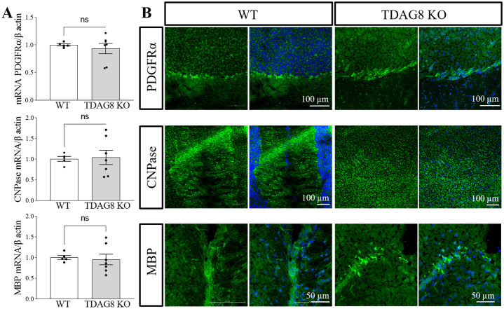
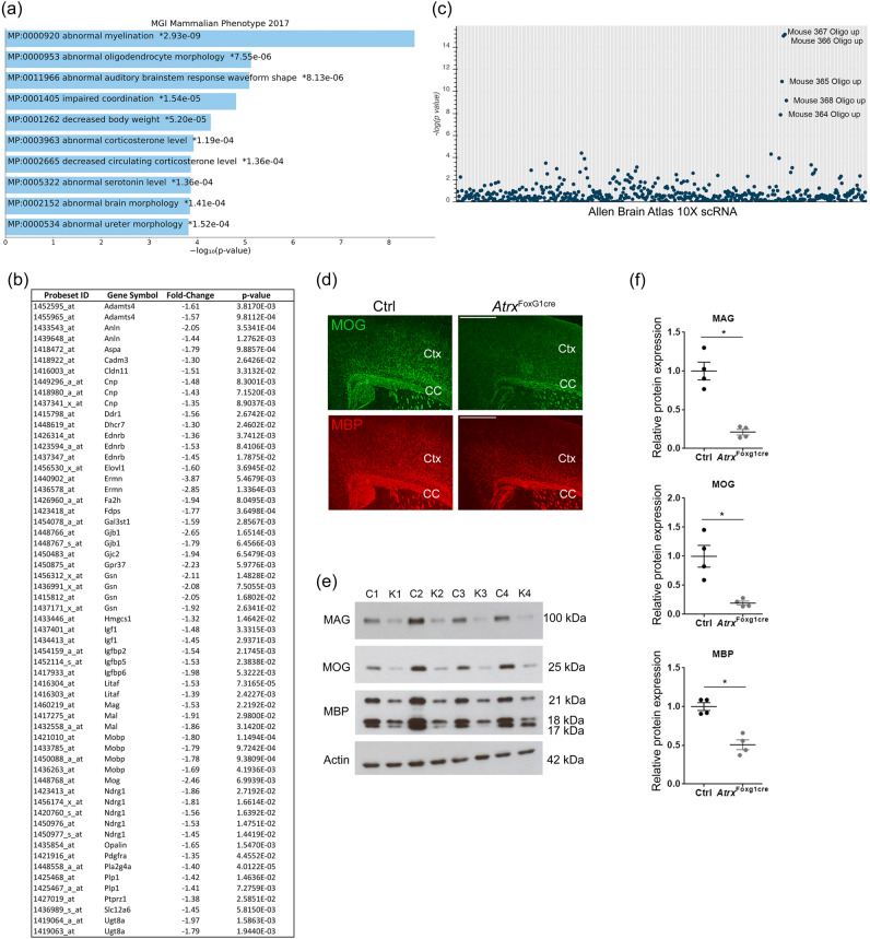
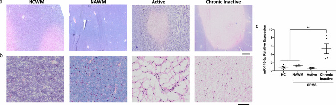
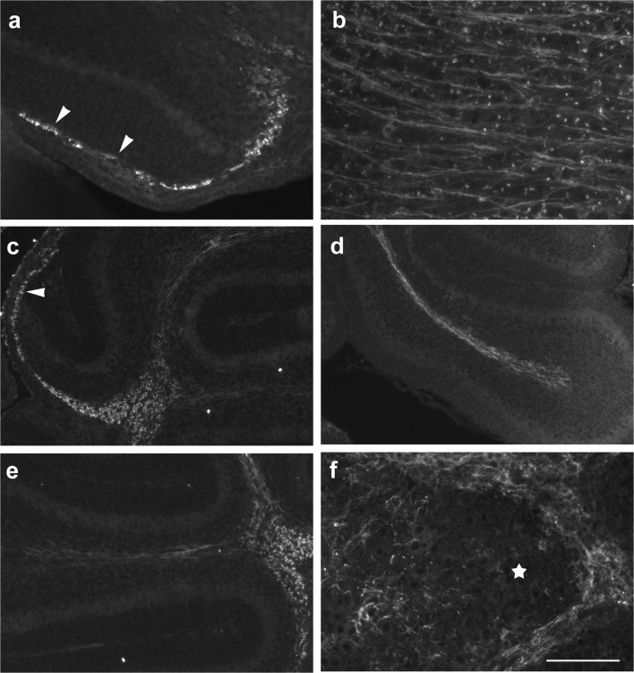
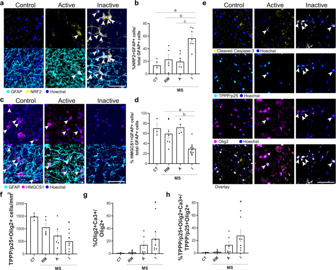
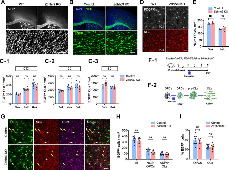
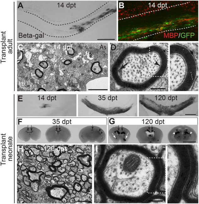
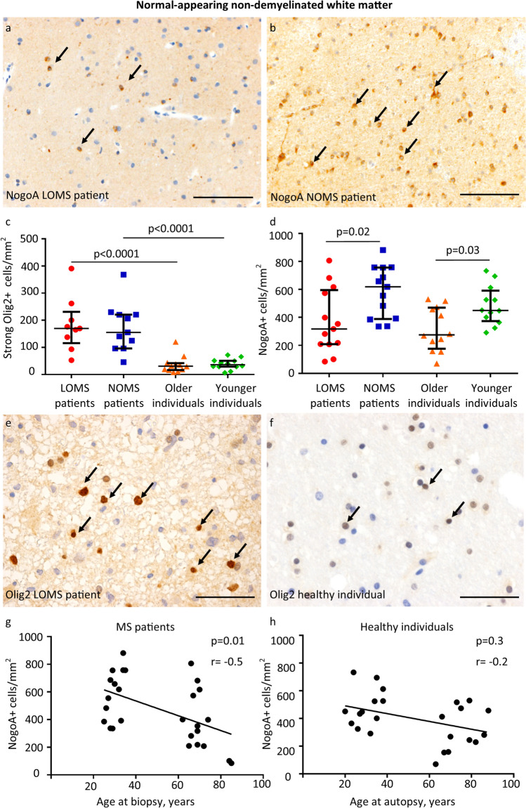
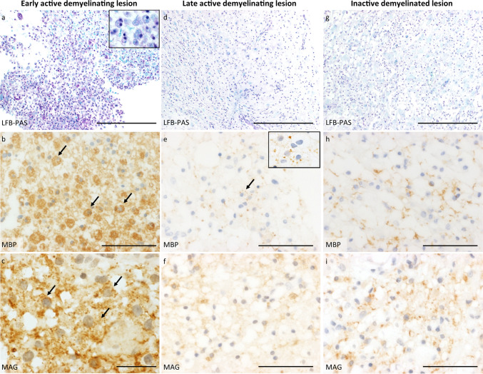
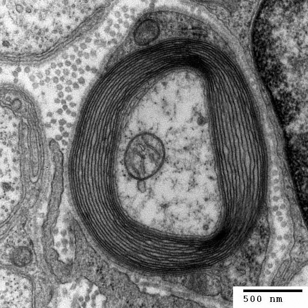
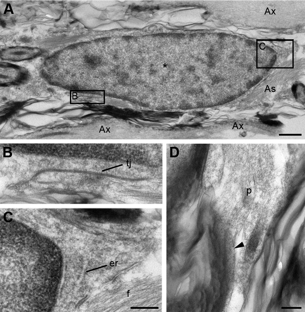
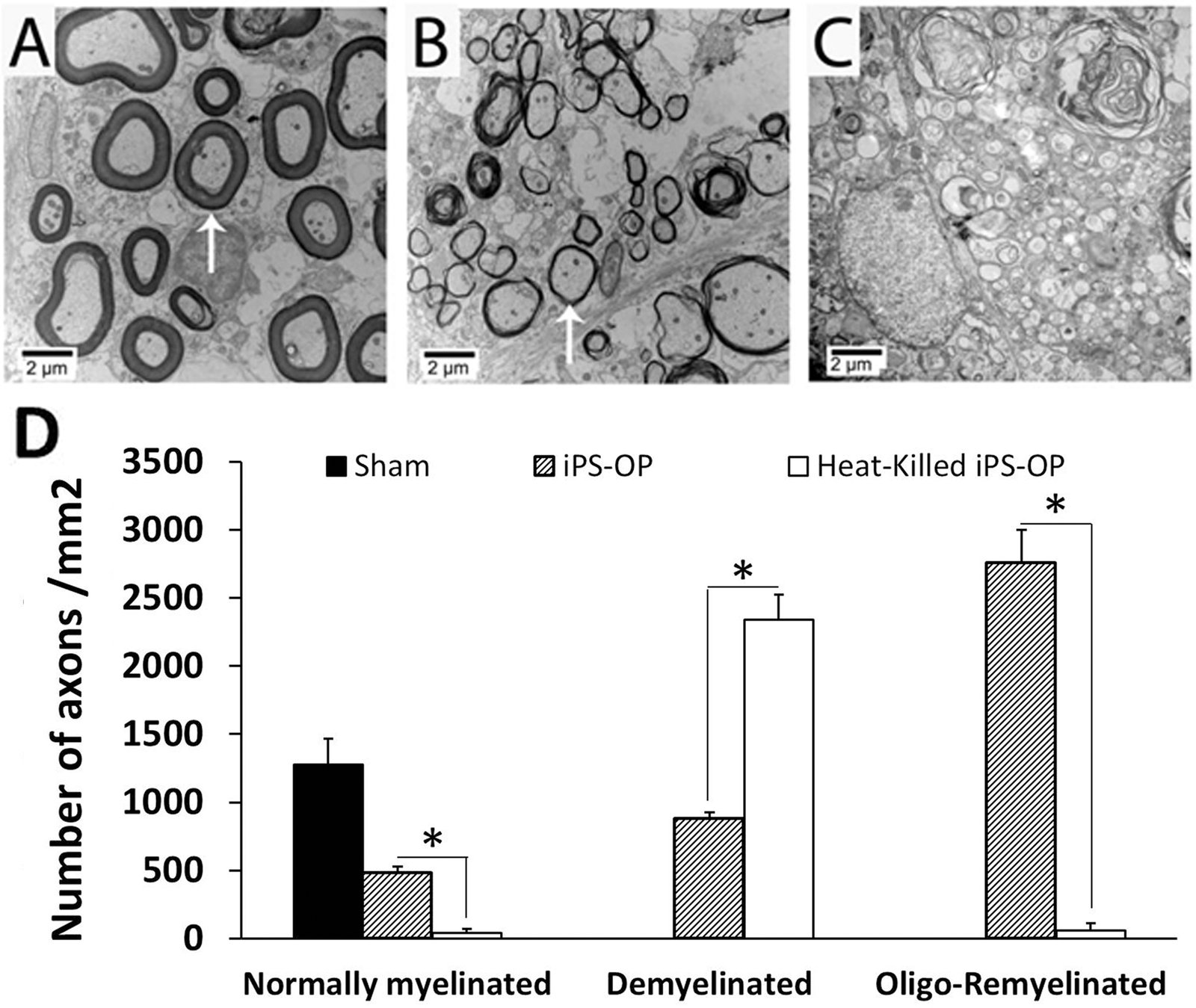
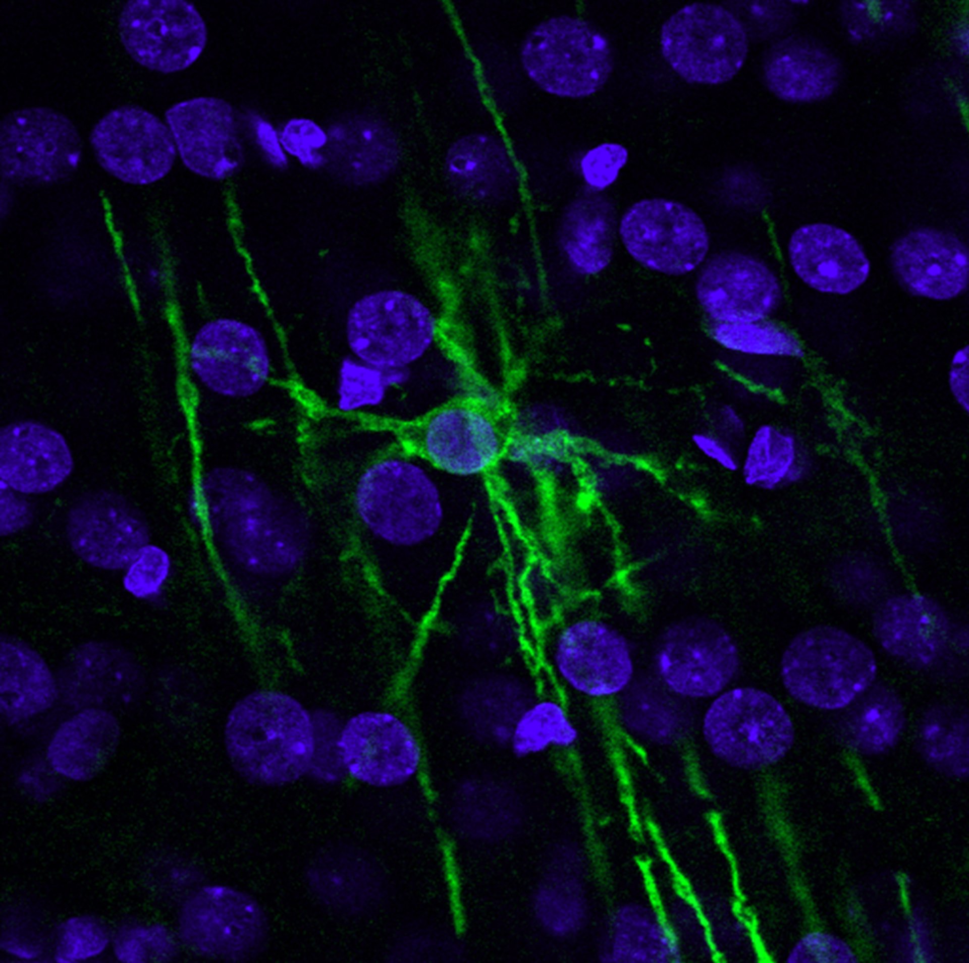
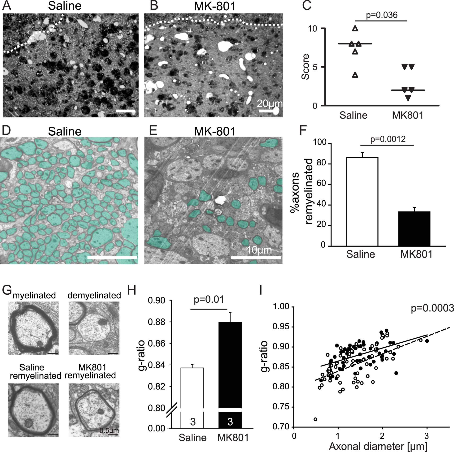
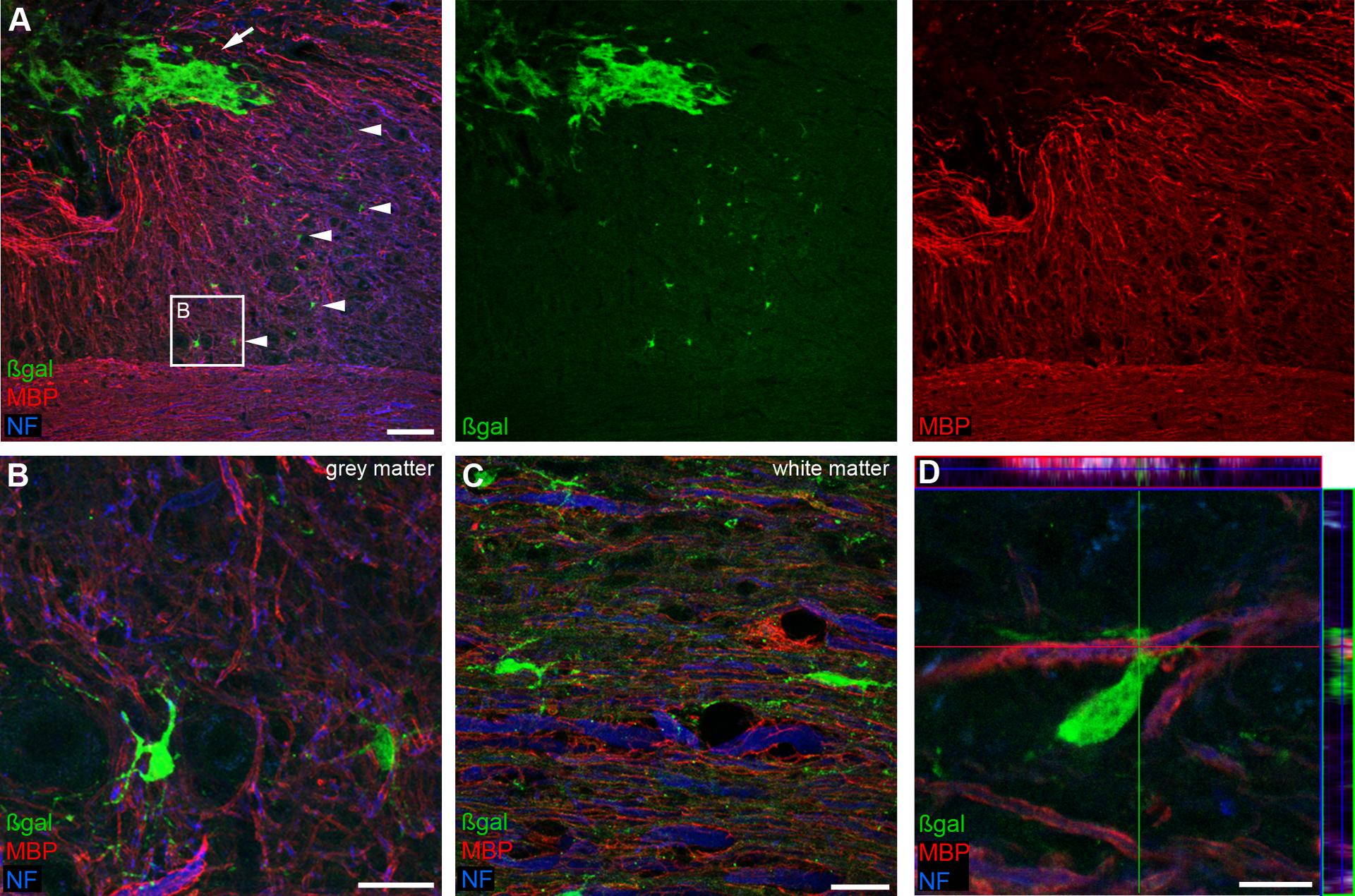
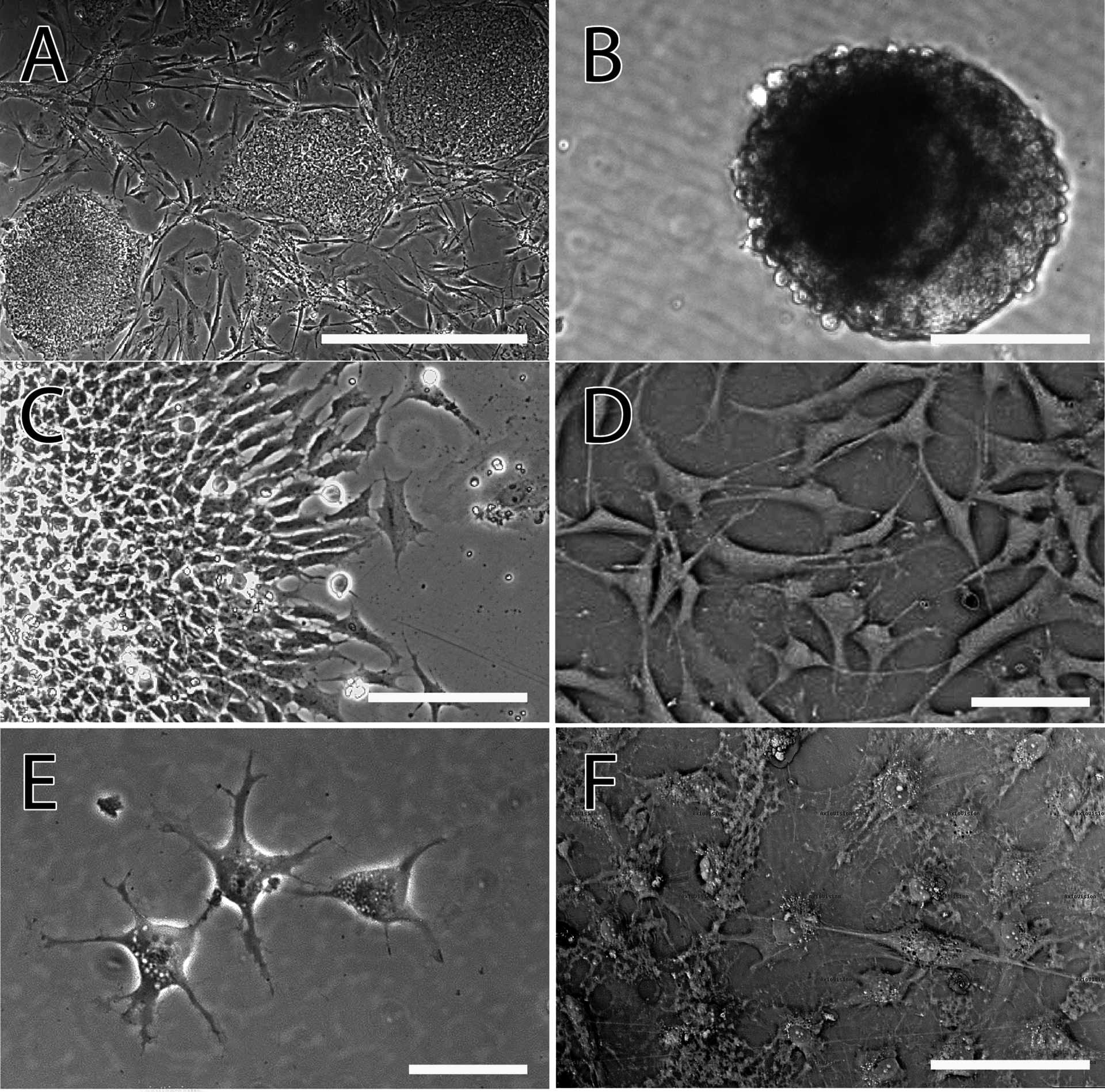
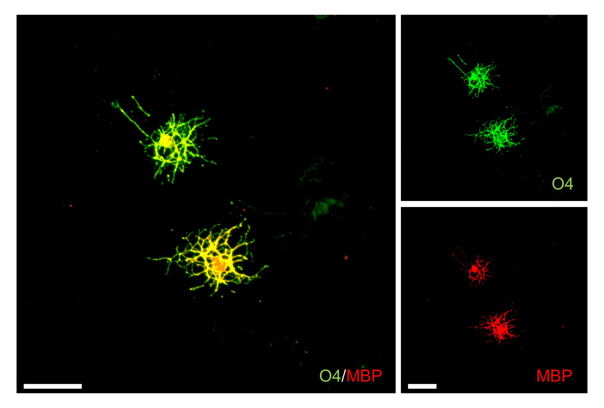
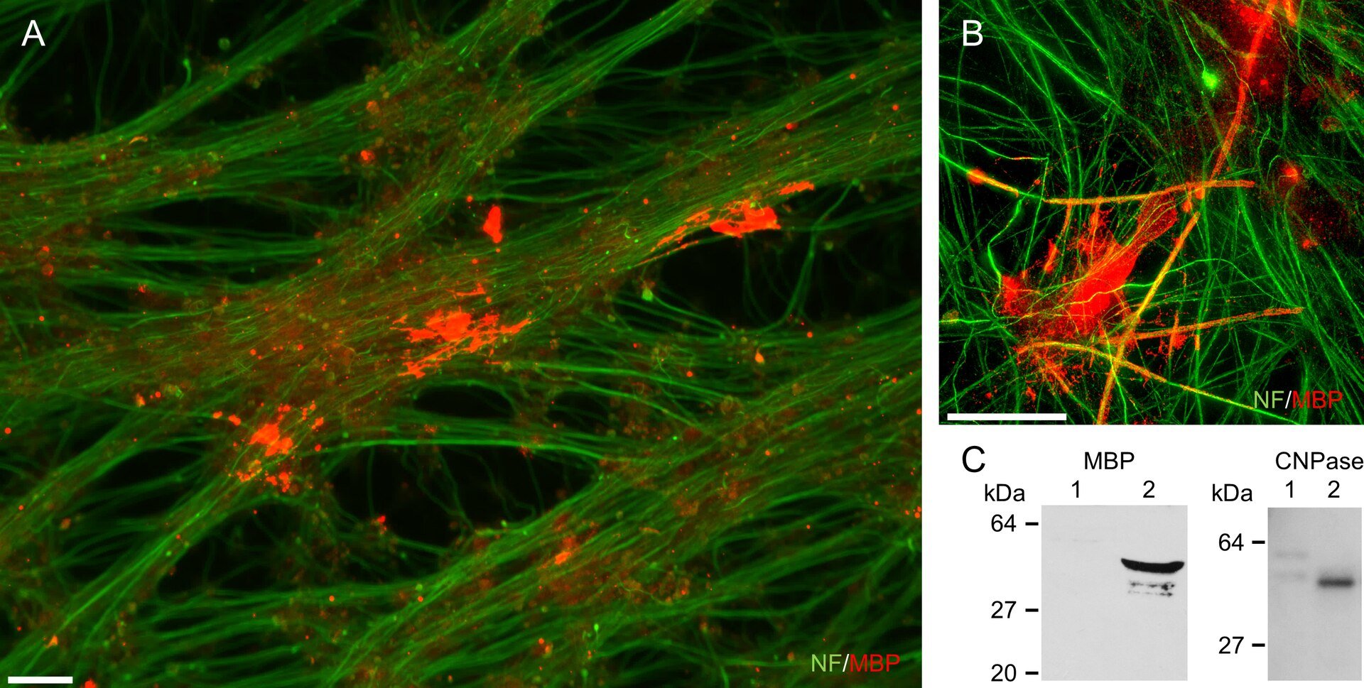
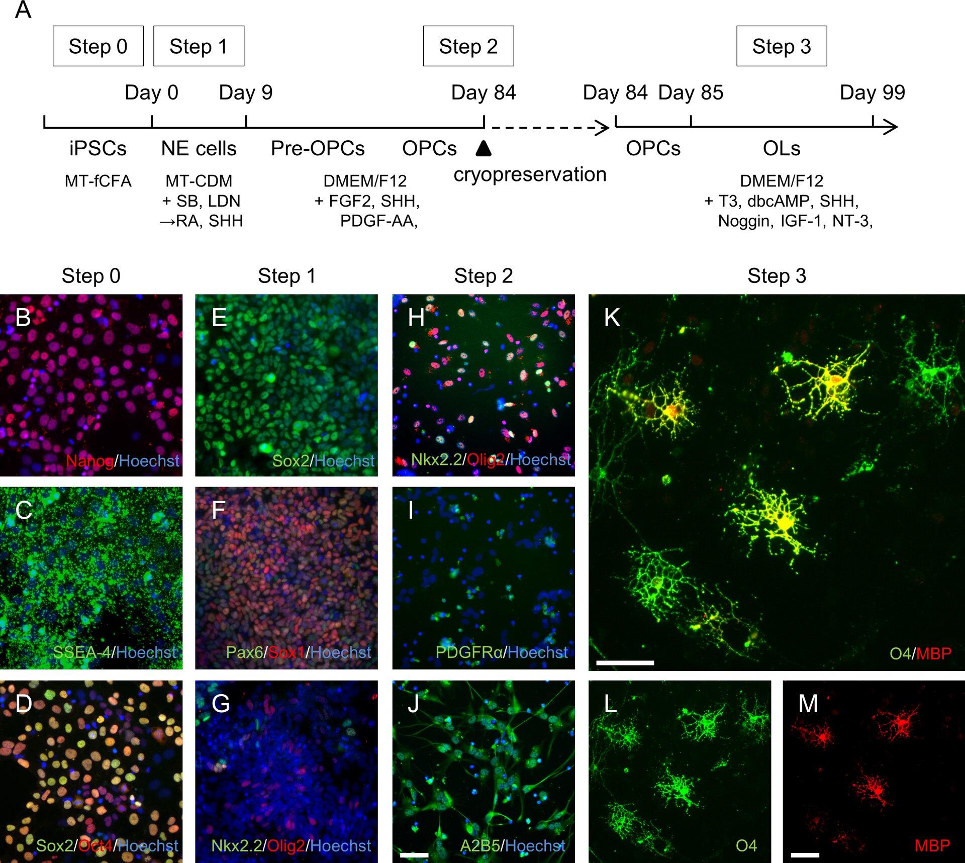
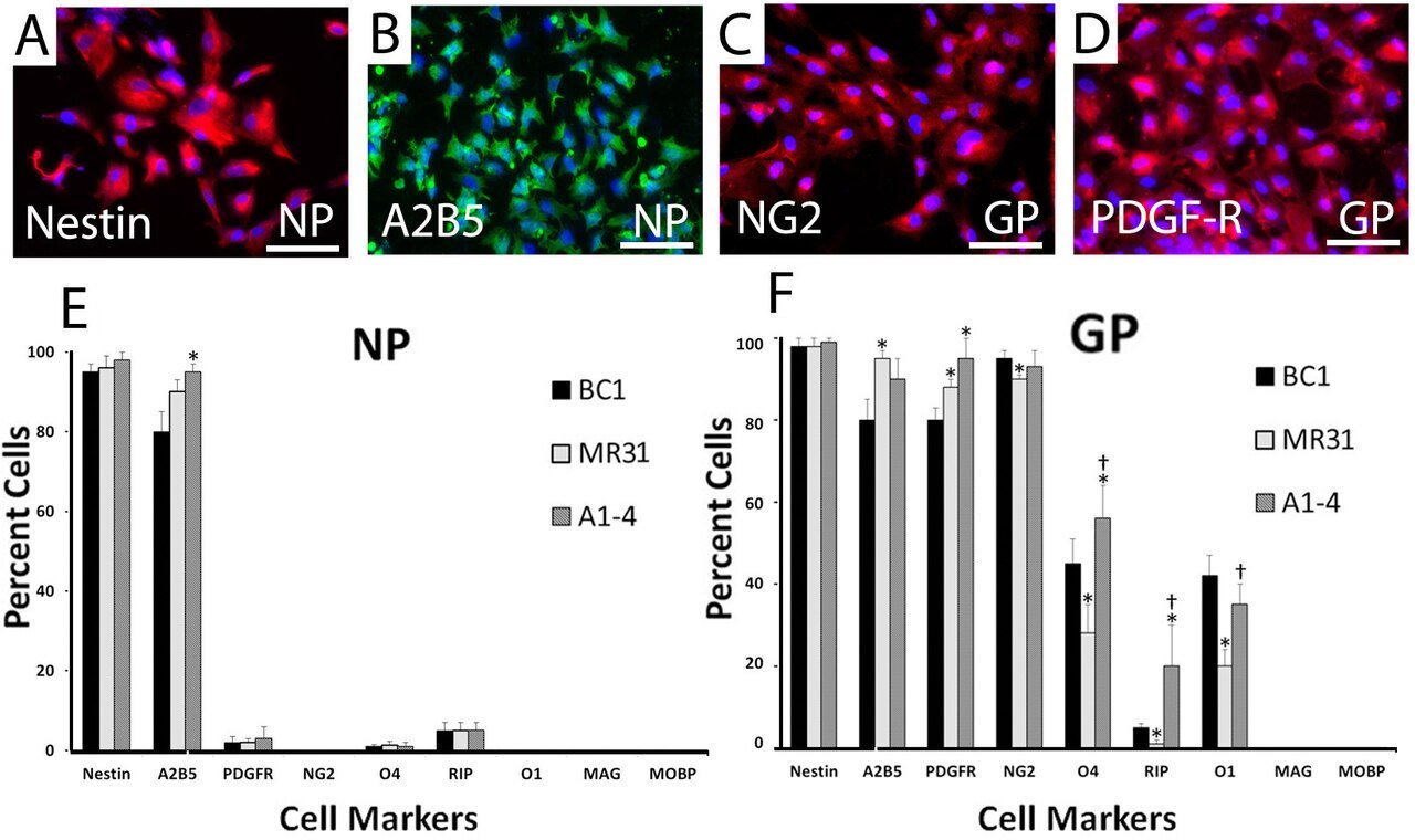
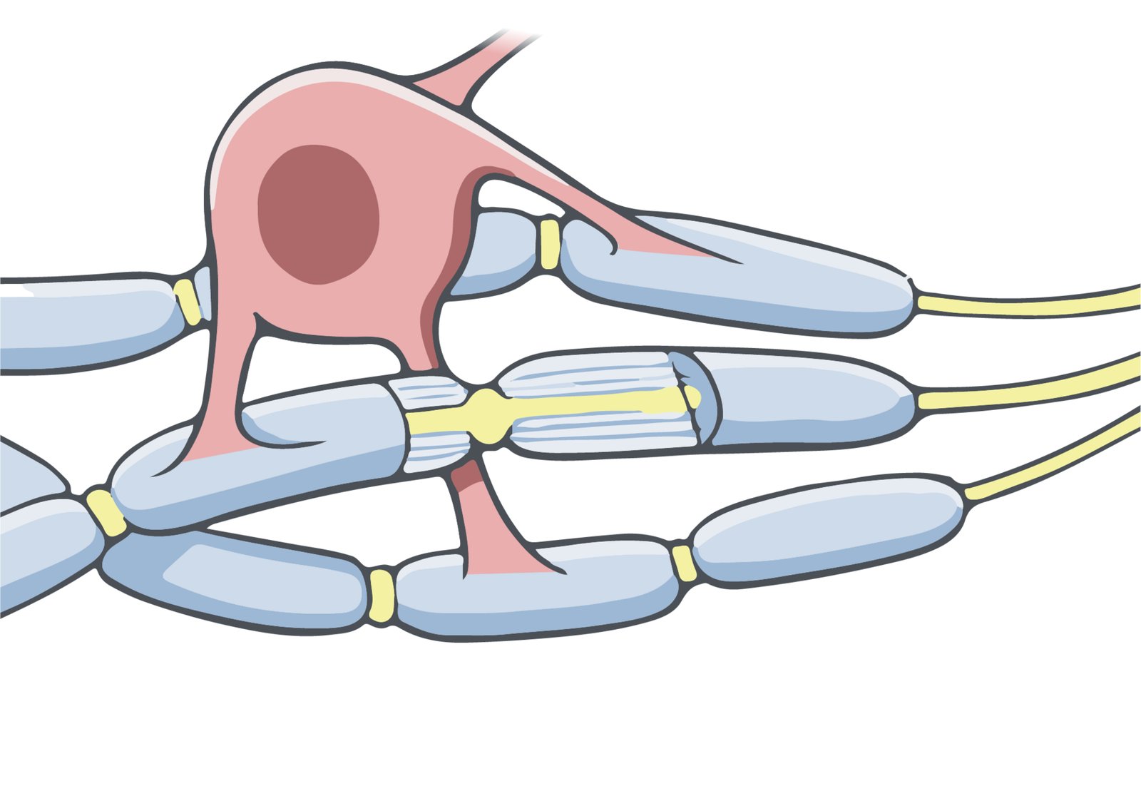
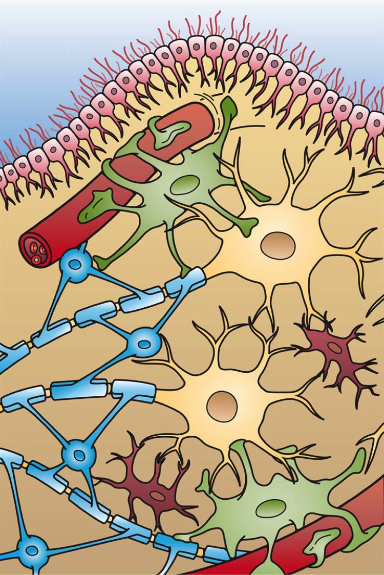
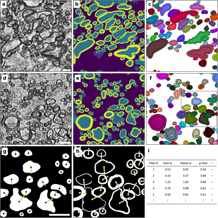
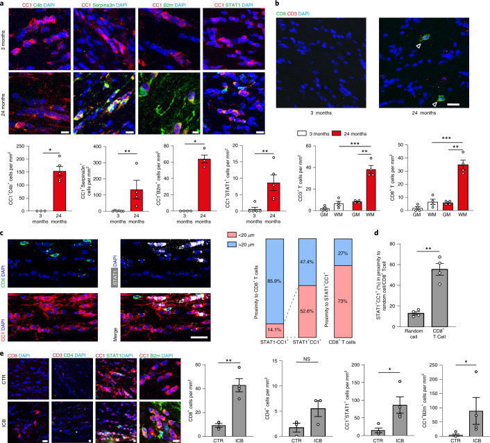
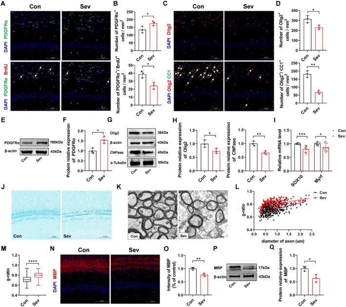
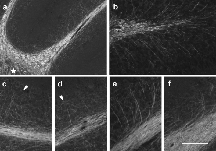
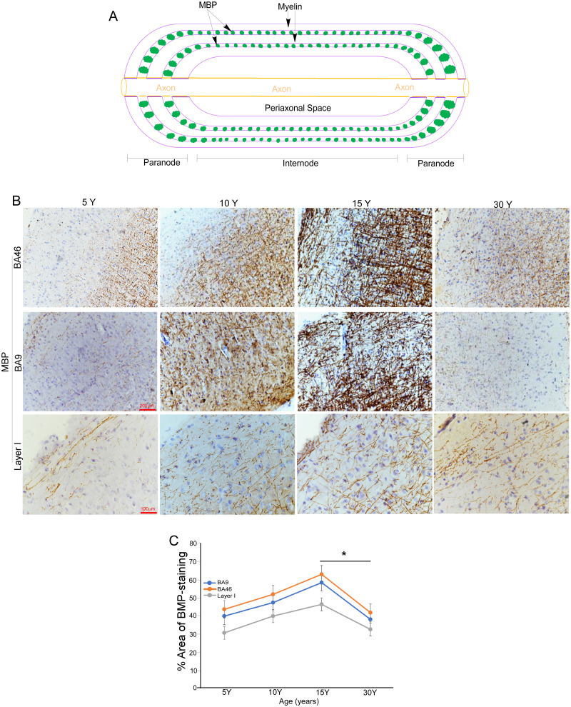
Transmission electron micrograph of a myelinated axon in transverse section. The concentric myelin lamellae are visible surrounding the axon core. The myelin sheath is produced by an oligodendrocyte in the CNS. · user:Roadnottaken (Wikimedia Commons)Electron micrograph of an ependyma-derived oligodendrocyte. From Meletis et al., showing the ultrastructure of an oligodendrocyte that arose from ependymal cells after spinal cord injury. · Meletis K, Barnabe-Heider F, Carlen M, Evergren E, Tomilin N et al. (Wikimedia Commons)TEM of transverse sections of rat spinal cord showing remyelination of spared axons by iPS-derived OPCs after contusive SCI. Thin remyelinating sheaths are visible around axons. · All AH, Gharibani P, Gupta S, Bazley FA, Pashai N, Chou BK et al. (Wikimedia Commons)Confocal image of oligodendrocytes in adult mouse brain stained with the Rip antibody (green). Cell nuclei are counterstained with DAPI (blue). The branching processes characteristic of myelinating oligodendrocytes are visible. · Oleg Tsupykov (Wikimedia Commons)Semithin sections of lesioned cerebellar white matter showing that remyelination is dependent on NMDA receptor activation. Remyelinating axons show thin myelin sheaths characteristic of newly formed oligodendrocyte internodes. · Lundgaard I, Luzhynskaya A, Stockley JH, Wang Z, Evans KA, Sibson N et al. (Wikimedia Commons)Ependymal cell-derived progeny after spinal cord injury. The image shows ependymal-derived cells immunoreactive for oligodendrocyte markers, demonstrating that ependymal cells can contribute oligodendrocytes to the injury response. · Meletis K, Barnabe-Heider F, Carlen M, Evergren E, Tomilin N et al. (Wikimedia Commons)Bright field microscopy showing the stages of oligodendrocyte differentiation from human iPSCs (BC1 line). The progression from OPC to immature and then mature oligodendrocyte can be followed by the elaboration of processes and flattening of the cell body. · All AH, Gharibani P, Gupta S, Bazley FA, Pashai N, Chou BK et al. (Wikimedia Commons)Immunofluorescence of oligodendrocytes differentiated from human iPSCs. O4 (green) marks the oligodendrocyte surface; MBP (red) marks myelin basic protein. The two markers co-localise in mature oligodendrocytes. · Yamashita T, Miyamoto Y, Bando Y, Ono T, Kobayashi S, Doi A et al. (Wikimedia Commons)Monkey ESC-derived OPCs (CMK6SFF) co-cultured with rat DRG neurons demonstrate myelinogenic potency in vitro. MBP-positive (red) myelin segments wrap along axons. · Yamashita T, Miyamoto Y, Bando Y, Ono T, Kobayashi S, Doi A et al. (Wikimedia Commons)OPCs and oligodendrocytes differentiated from human iPSCs (A3 line). Fluorescence images show the differentiation protocol outcome and marker expression. · Yamashita T, Miyamoto Y, Bando Y, Ono T, Kobayashi S, Doi A et al. (Wikimedia Commons)Indirect immunofluorescence defining the early stages of oligodendrocyte differentiation from iPSCs into neural progenitors (NP) and glial progenitors (GP). · All AH, Gharibani P, Gupta S, Bazley FA, Pashai N, Chou BK et al. (Wikimedia Commons)NIH BioArt illustration of an oligodendrocyte showing its branching processes, which wrap around multiple axons to form the myelin sheath. Public domain image courtesy of NIAID. · Courtesy of NIAID / Ryan Kissinger (Wikimedia Commons)Scientific illustration showing the four types of glial cells in the CNS: ependymal cells, astrocytes, microglia, and oligodendrocytes. The oligodendrocyte is shown with processes wrapping around axons. · Artwork by Holly Fischer (Wikimedia Commons)Overview of the segmentation approach for electron microscopic data. a, Preprocessed transmission electron microscopic data of human corpus callosum (CC). b, Semantic prediction of the preprocessed data by the trained DenseNet, returning a likelihood of each pixel to belong to either background (violet), axon (teal), or myelin (yellow). c, Post-processed final segmentation. Each instance of a fiber (axon and myelin sheath) is labelled with a different random color. d–f, Analogous to a–c, with transmission electron microscopic data of human superficial white matter (SWM). The dashed region in f is shown in g,h. The measurement of axon (g) and myelination (h) diameters is based on ellipses fitted to each structure. Points show the centroid of each structure, axes show half of the minor axis of the fitted ellipse for the axon (solid line) and myelin (dashed line). i, Measurements (SWM: n = 220,431, CC: n = 163,133) from g, h are used for statistical assessment of local ultrastructure. We treat the short axis (half of which is shown in yellow in g, h) of the fitted ellipse as the actual diameter of each structure. All scale bars are 2 µm. · Ruthig, von der Planitz, Morozova et al 2025, PLOS BiologyIFN-responsive oligodendrocytes localize to aged white matter close to CD8+ T cells. a, Immunofluorescence staining and quantification of C4b, Serpina3n, B2m and STAT1 in CC1+ oligodendrocytes in the white matter of 3- and 24-month-old mice (C4b+CC1+, 3-month, n = 3, 24-month, n = 5; Serpina3n+CC1+, 3-month, n = 6, 24-month, n = 4; B2m+CC1+, 3-month, n = 4, 24-month, n = 4; STAT1+CC1+, 3-month, n = 5, 24-month, n = 5; data are mean ± s.e.m.). Scale bar, 20 µm; for B2m, 10 µm. · Kaya, Mattugini, Liu et al 2022, Nature NeuroscienceEffects of repeated sevoflurane exposure on OPC proliferation, differentiation, and myelination in neonatal mice. (A) IF staining of PDGFRα (green) and BrdU (red) in the hippocampal CA1 region. Colocalized cells (yellow) are indicated by white arrows, and nuclei were counterstained with DAPI (blue) (scale bar = 100 μm). (B) Quantitative analysis of PDGFRα+ cells and PDGFRα+/BrdU+ double-positive cells; n = 3 mice/group. (C) IF staining of Olig2 (red) and CC1 (green) in the hippocampal CA1 region. Colocalized cells (yellow) are indicated by white arrows, and nuclei were counterstained with DAPI (blue) (scale bar = 100 μm). (D) Quantitative analysis of Olig2+ cells and Olig2+/CC1+ double-positive cells; n = 3 mice/group. (E) WB analysis of PDGFRα protein expression in hippocampal tissue. (F) Quantitative analysis of PDGFRα protein band intensity; n = 3 mice/group. (G) WB analysis of Olig2 and CNPase protein expression in hippocampal tissue. · Liu, Bai, Liu et al 2026, CNS Neuroscience & TherapeuticsMBP immunoreactivity in the p15 and p28 cerebellum. a–d The p15 cerebellum is characterized by intensive immunoreactivity for MBP throughout the WM. The density of MBP-positive fibers also increased within the DCN (star in a) and the IGL of all lobules. In the proximal parts of the lobules, MBP-positive fibers are found almost up to the PCL (a, c). Distally, MBP-positive fibers do not yet reach the PCL (b, d). At the tips of the lobular WM, MBP-positive fibers branch radially in all directions (b). Arrowheads in c and d point out MBP-positive fibers running more or less parallel to the PCL. e, f In the p28 cerebellum, many MBP-positive fibers ascend through the IGL up to the PCL. Whereas they emanate at flat angles from the white matter in proximal parts of the lobules (e), more distally, · Groteklaes, Bönisch, Eiberger et al 2020, Cerebellum (London, England)Age-related changes in the intensity of MBP signals in the macaque brain. A, Schematic diagram illustrates the role of MBP in maintaining myelin sheath integrity. B, Representative images of immunohistochemistry (IHC) showing MBP signals (brown) in Brodmann areas BA46 and BA9, as well as layer 1 of the prefrontal cortex (PFC) in macaques at ages 5, 10, 15, and 30 years. Scale bar as indicated. C, Relative intensities of IHC MBP signals shown in panel B were quantified using ImageJ. Data are presented as mean ± SD (n = 5 macaques per age group; * p = 0.0038, unpaired t test). · Zhang, Yao, Lv et al 2026, eNeuroProphylactic OM-MOG prevents the development of spinal cord neuropathology during MOG-EAE in DR2b.Ab° mice. Neuropathological analysis of spinal cord sections from prophylactic vehicle- (upper and middle panels) and OM-MOG-injected (lower panels) DR2b.Ab° mice on day 36 post-immunization for EAE. Inflammatory cell infiltration was visualized by H&E (A, D); demyelination by Luxol fast blue [(B) and enlarged inset, (E)]; axon damage by Bielschowsky's silver staining [(C) and enlarged inset, (F)]. Vehicle-treated mice show large confluent inflammatory, demyelinating lesions with axon damage typical of MOG-EAE (arrowheads). OM-MOG-vaccinated mice showed no spinal cord pathology. Scale bars 500 μM (A–F), 100 μM enlarged inserts in middle panels. · Dagkonaki, Avloniti, Evangelidou et al 2020, Frontiers in ImmunologyTDAG8 deficiency does not affect normal myelination in the mouse brain. A. The mRNA analysis of pdgfrα, cnpase and mbp expression in whole brain homogenates did not reveal differences between the TDAG8 KO and WT mice. B. Representative images of immunohistochemically stained WT and TDAG8 KO mice brains (cerebellum) show no differences in myelination between the two genotypes. · Caratis, Opiełka, Hausmann et al 2024, PLOS ONEDefective myelination in the ATRX-null mouse forebrain. d Immunofluorescence microscopy of P20 brain cryosections stained with anti-MOG (green) and anti-MBP (red) antibodies confirms decreased levels of these myelin proteins in the cortex (Ctx) and corpus callosum (CC) of the forebrain of AtrxFoxG1Cre mice compared to controls (Ctrl) (n = 3 animals for each genotype). · Rowland, Jiang, Shafiq et al 2023, Nature CommunicationsmiR-145-5p is upregulated in chronic lesion tissue from SPMS brains. a, b Luxol fast blue/hematoxylin and eosin stained human brain tissue from healthy control white matter (HCWM), and normal appearing white matter (NAWM), active and chronic inactive lesion tissue from SPMS brain. a Scale bar = 1 mm. b Magnified from (a); scale bar = 50 µm. · Kornfeld, Cummings, Yaworski et al 2024, Communications BiologyCNP-1 immunoreactivity in the early postnatal mouse cerebellum (p0–p6). a CNP-1-positive cells in the p4 cerebellar anlage are roundish and may be found in the velum medullare (arrowheads in a) and the anteriormost parts of the cerebellar anlage. Sagittal section close to the midline; anterior is to the left. b In the brainstem, CNP-1-positive structures have a fibrous morphology at this age. c, d CNP-1-positive cells at p6. Immunoreactive cells can again be detected in the velum medullare (arrowhead in c). They can now also be seen in the nascent central WM and the nascent WM extending into t · Groteklaes, Bönisch, Eiberger et al 2020, Cerebellum (London, England)Astrocytic Nrf2 and cholesterol pathways are altered in chronic human brain lesions with poor remyelination potential and oligodendrocyte death. a Nrf2+ (yellow) astrocytes (GFAP+; cyan) (arrows) with Hoechst in blue. Scale bar, 100 μm. e Active caspase-3+ (yellow) oligodendrocytes (TPPP/p25+; cyan, and Olig2+; magenta) (arrows) in control (CT), remyelinated (RM), active (A) and inactive lesions (I) from multiple sclerosis (MS) cases. · Molina-Gonzalez, Holloway, Jiwaji et al 2023, Nature CommunicationsNo detectable gross abnormality in oligodendrocyte development in Zdhhc9 KO mice. ( A ) Fluorescent (upper) an · Jeong, Gonzalez-Fernandez, Crawley et al 2025, eLifeNeural precursors form compact myelin sheaths after transplantation into adult or neonatal Plp1-tg mice. ( A ) Light microscopy image of beta galactosidase-positive cells in the corpus callosum (delineated) of a Plp1-tg transplant recipient, 14 days post transplantation. ( B ) Corpus callosum of another transplant recipient showing that transplanted GFP-positive neurospheres form MBP-positive myelin. ( C and D ) Electron micrographs showing that the transplanted cells generate oligodendrocytes that wrap axons with normal-appearing myelin sheaths in which the inner tongue (black arrow), periaxonal space (arrowhead) · Gruenenfelder, McLaughlin, Griffiths et al 2020, BrainAnti-Olig2 staining of the white matter of a healthy older individual: Both strongly (arrows) and weakly Olig2-positive nuclei are identified. · Stork L, Stephan J, Kutllovci A et al. Impaired remyelination in late-onset multiple sclerosis. Acta Neuropathol. 2025. PMID 40167776Histopathology of different demyelinating lesion activities. Panels a-c illustrate an early active demyelinating lesion, representing the initial stage of demyelination. At this stage, degradation products from both major (e.g., MBP) and minor (e.g., MAG) myelin proteins are present within phagocytes. a LFB/PAS staining reveals a demyelinated lesion containing multiple foamy macrophages. The inset shows an enlarged microscopic image of foamy macrophages with LFB-positive myelin debris; b Anti-myelin basic protein (MBP) staining of the early active lesion reflects myelin destruction and highlights numerous macrophages filled with myelin degradation products; c Anti-myelin-associated glycoprotein (MAG) staining also shows multiple macrophages containing MAG-positive degradation products, a minor myelin protein. Panels d-f depict a late active demyelinating lesion, representing the subsequent stage of demyelination, where only degradation products from major myelin proteins (e.g., MBP) can be identified within phagocytes. d LFB/PAS staining shows a completely demyelinated lesion with reactive astrogliosis and foamy macrophages; e Anti-MBP staining reflects an advanced stage of demyelination, with only a few macrophages containing MBP-positive degradation products (see inset for MBP-positive macrophages); f In contrast, no degradation products from minor myelin proteins, such as MAG, are detected within macrophages, as demonstrated by anti-MAG staining. Panels g-i present an inactive demyelinated lesion, where no active demyelination processes are observed. g LFB/PAS staining reveals a demyelinated lesion accompanied by reactive astrogliosis and foamy macrophages; h Anti-MBP staining shows newly formed, thin myelinated fibers, indicating early remyelination. However, no myelin degradation products are observed within macrophages; i Anti-MAG staining also identifies early remyelination. LFB/PAS Luxol fast blue/periodic acid-Schiff, MBP myelin basic protein, MAG myelin-associated glycoprotein, Scale bars: a, d and g 200 um; b, f and i 50 um; e and h 100 um; c 20 um · Stork, Stephan, Kutllovci et al 2025, Acta Neuropathologica
Oligodendrocyte knowledge base
Maintained by Oligodendro. Cite or omit. No em dashes. Last foundation pass: 2026-09-09.Citation audit 2026-09-09: rows carrying a PMID were resolved through NCBI and their titles checked against the claim. Rows without a PMID are textbook or review attributions that have not yet been resolved to a PubMed record; treat those as thin until they are.
History of discovery
The cell was named for its shape before anyone knew what it did. Sixty years separated the name from the proof of function.
Ramon y Cajal first described cells in CNS white matter distinct from neurons and astrocytes, calling them "third element" cells.
Ramon y Cajal, Histologie du Systeme Nerveux, 1909
Pio del Rio-Hortega used a silver carbonate method in 1921 to distinguish two populations of non-neuronal CNS cells: microglia and oligodendroglia. He named oligodendrocytes for their few (oligo) branching processes (dendro).
Rio-Hortega, Mem Real Soc Esp Hist Nat, 1921
The myelin sheath had been described by Virchow in 1854 as a lipid-rich insulating material, but its cellular origin in the CNS was not confirmed until electron microscopy in the 1960s linked oligodendrocyte processes to compact myelin lamellae.
Peters, Journal of Biophysical and Biochemical Cytology, 1960
The discovery that one oligodendrocyte myelinates multiple axons (unlike the one-to-one Schwann cell relationship in the PNS) was established by EM serial reconstruction.
Bunge et al., Journal of Biophysical and Biochemical Cytology, 1961
Function
Insulation is the famous job and not the only one. The cell also feeds the axon it wraps, and that second role is separable from the first.
Myelination increases axonal conduction velocity up to 100-fold by enabling saltatory conduction and reduces the metabolic cost of action potential propagation.
Hartline & Colman, Current Biology, 2007
Myelin compaction reduces membrane capacitance and prevents ion leak between nodes, which together account for the velocity and efficiency gains of saltatory conduction.
Hartline & Colman, Current Biology, 2007
Oligodendrocytes provide metabolic support to axons by delivering lactate and pyruvate via monocarboxylate transporters (MCT1 in oligodendrocytes, MCT2 in axons), independent of myelination. Loss of this support causes axon degeneration even when myelin appears intact.
One cell body, many arms, each arm ending in a separate sheath on a separate axon. That reach is what distinguishes it from its peripheral counterpart.
A single mature CNS oligodendrocyte extends processes to myelinate up to ~50 distinct axon internodes; one Schwann cell wraps exactly one internode.
On H&E, mature oligodendrocytes show a round, dark nucleus surrounded by a clear perinuclear halo, a formalin fixation artifact from cytoplasmic retraction. The halo disappears in frozen or glutaraldehyde-fixed tissue.
Bancroft & Gamble, Theory and Practice of Histological Techniques, 6th ed.
Schmidt-Lanterman incisures are cytoplasm-filled clefts in compact myelin that provide a conduit for metabolite exchange between the oligodendrocyte soma and the inner tongue.
Ghabriel & Allt, Brain Research, 1981
Structure: the sheath
Compact myelin is mostly lipid, held together by two proteins, and interrupted at regular intervals so the impulse can be regenerated.
Myelin is a multilamellar lipid-rich membrane (70% lipid, 30% protein by dry weight) compacted by myelin basic protein (MBP) and proteolipid protein (PLP), which together account for ~80% of myelin protein.
The internode is the myelin-covered axon segment between two nodes of Ranvier. Internode length scales with axon diameter; larger axons have longer internodes.
Waxman & Bennett, Nature New Biology, 1972
Nodes of Ranvier are bare axon segments (~1 micron) flanked by paranodes and juxtaparanodes; voltage-gated Nav1.6 channels cluster at the node to enable saltatory conduction.
Precursors arrive in waves from the ventricular zone and are identified by a marker pair that is lost the moment they mature.
Oligodendrocyte precursor cells (OPCs, also called O-2A progenitors) arise in three successive waves from the ventral and then dorsal ventricular zone progenitors during embryogenesis.
Richardson et al., Nature Reviews Neuroscience, 2006
OPCs are identified by co-expression of NG2 (CSPG4) and PDGFRalpha; both markers are downregulated upon differentiation to mature oligodendrocytes.
The transcription factors Olig1 and Olig2 are expressed throughout the oligodendrocyte lineage from OPC to mature cell; Olig2 is the standard IHC marker for the full lineage.
Maturation is held back as much as it is pushed forward, and precursors persist into adult life rather than being spent during development.
OPC differentiation to myelinating oligodendrocyte is promoted by PDGF-A, FGF2, and thyroid hormone, and inhibited by Notch signalling and PSA-NCAM on axons.
Barres & Raff, Journal of Cell Biology, 1994
OPCs persist throughout adult white and grey matter as the principal source of remyelination after injury, retaining differentiation capacity across the lifespan.
The transcriptional repressor YY1 and chromatin remodelling by histone deacetylases are required for OPC-to-oligodendrocyte transition; HDAC inhibition blocks differentiation.
He et al., Nature Neuroscience, 2007
Myelination biology
The axon, not the oligodendrocyte, decides whether it is wrapped and how thickly. Wrapping then proceeds from the inside out.
Axonal signals govern whether an OPC myelinates a given axon; minimum axon diameter for CNS myelination is ~0.2 microns. Below this threshold, axons remain unmyelinated.
Voyvodic, Journal of Neuroscience, 1989
Neuregulin-1 (NRG1) on the axon surface signals via ErbB receptors on OPCs to regulate the onset and extent of myelination.
Myelin assembly begins at the inner tongue, which spirals around the axon; compaction proceeds outward as MBP displaces cytoplasm from the growing sheath layers.
The disease that made this cell clinically important. Myelin is the target; the axon is what is ultimately lost.
In multiple sclerosis, autoimmune T-cell and antibody-mediated attack targets myelin and oligodendrocytes, producing plaques with LFB pallor and relative early axon preservation.
Compston & Coles, Lancet, 2008
MS plaques are classified histologically as active (macrophages with myelin debris), chronic active (active rim, inactive centre), and inactive (gliotic scar, no macrophages).
Lassmann et al., Lancet Neurology, 2012
Shadow plaques are areas of incomplete or thin remyelination visible as pale LFB staining adjacent to a fully demyelinated plaque; they indicate endogenous repair.
Prineas & Connell, Annals of Neurology, 1979
Axonal injury in MS occurs from the earliest stages and is the main substrate of permanent disability; it can occur in demyelinated and in myelinated axons.
Pathology: genetic and infectious white matter disease
Myelin also fails without autoimmunity: by inherited defect in its synthesis, by attack on the neighbouring astrocyte, and by a virus that infects the oligodendrocyte directly.
Leukodystrophies are genetic disorders of myelin synthesis or maintenance; most involve mutations in oligodendrocyte-expressed genes (PLP1 in PMD, GALC in Krabbe, ARSA in MLD).
Vanderver et al., Journal of Child Neurology, 2015
Neuromyelitis optica spectrum disorder (NMOSD) is associated with aquaporin-4 antibodies targeting astrocytes, but causes severe oligodendrocyte and myelin loss secondarily in spinal cord and optic nerves.
Pittock & Lucchinetti, Annals of Neurology, 2016
Progressive multifocal leukoencephalopathy (PML) is caused by JC virus replication in oligodendrocytes, producing widespread CNS demyelination in immunocompromised patients.
Berger, Annals of Neurology, 2011
Disease models
Three preparations, each isolating a different mechanism. Choosing the wrong one is the commonest way to misread a remyelination result.
Cuprizone (dietary copper chelator) selectively kills mature oligodendrocytes in mice, producing reproducible corpus callosum demyelination and the dominant preclinical platform for remyelination kinetics. It does not replicate the immune-mediated lesion of MS.
EAE (experimental autoimmune encephalomyelitis) is induced by immunisation with myelin peptides (MOG35-55, MBP, PLP) and models immune-mediated demyelination; cuprizone models toxic-metabolic demyelination without significant T-cell infiltration.
Ransohoff, Nature Reviews Immunology, 2012
Lysolecithin (lysophosphatidylcholine) focal injection produces rapid, well-demarcated demyelinating lesions and spontaneous remyelination within weeks; useful for studying local repair mechanisms.
Jeffery & Blakemore, Journal of Neurocytology, 1995
Remyelination
The CNS can rebuild myelin, and the repair is recognisable because it is thinner than the original. Where repair fails, the precursors are usually present but stalled.
Remyelination in the adult CNS is mediated primarily by surviving OPCs that are recruited to lesions, proliferate, and redifferentiate.
Remyelinated axons show thinner sheaths relative to axon diameter (elevated g-ratio compared to original myelin), a quantifiable marker of repair rather than regeneration.
Remyelination restores saltatory conduction and provides trophic support to axons, preventing secondary axon degeneration in chronically demyelinated lesions.
Irvine & Bhatt, Journal of Neurology, 2006
Remyelination failure in progressive MS is associated with OPC differentiation arrest, not absence of OPCs; OPCs are present in chronic lesions but do not mature.
Retinoid X receptor gamma (RXRgamma) agonists and antimuscarinics (clemastine, benztropine) promote OPC differentiation and remyelination in rodent models.
Myelin is stained for its lipid. Every white matter workup starts with one of these and lives or dies on the differentiation step.
Luxol fast blue (LFB) stains intact myelin phospholipids blue; pallor indicates demyelination and is the histological anchor for every white-matter differential diagnosis. Differentiation in lithium carbonate is critical: under-differentiation leaves artefactual background blue; over-differentiation washes out true myelin signal.
Bancroft & Gamble, Theory and Practice of Histological Techniques, 6th ed.
Kluver-Barrera combines LFB with cresyl violet: myelin stains blue, Nissl substance (neuronal rough ER) stains purple. Standard for evaluating both myelin integrity and neuronal architecture on a single section.
Kluver & Barrera, Journal of Neuropathology and Experimental Neurology, 1953
Paired LFB and Bielschowsky silver staining distinguishes primary demyelination (myelin lost, axons preserved on Bielschowsky) from axonal loss (both myelin and axon cylinders absent).
Weller, Brain Pathology, 1998
Methods: immunohistochemistry and electron microscopy
Antibodies find the protein before the lipid stain notices anything is wrong. Electron microscopy remains the arbiter of sheath thickness.
MBP immunohistochemistry is more sensitive than LFB for early or patchy demyelination and confirms myelin sheath integrity at the protein level. Loss of MBP signal precedes visible LFB pallor in early lesions.
Lassmann, Brain Pathology, 1998
MOG (myelin oligodendrocyte glycoprotein) and CNPase IHC mark the outer myelin membrane and oligodendrocyte cytoplasm respectively; useful for identifying remyelinating sheaths (thin MOG+ rings) and mature oligodendrocyte cell bodies (CNPase+).
Brunner et al., Journal of Neurochemistry, 1989
Electron microscopy remains the gold standard for measuring g-ratio and confirming myelin compaction, but confocal quantification of fluorescence ratios (MBP/NF200) provides a practical research alternative.
Four that changed what the cell is understood to do, and what its failure costs the axon.
Paper Barres, Raff et al. 1994: control of oligodendrocyte number, and the hormonal timing of their development ·Why it matters Developmental framework
The last decade reframed the cell twice: as a family of transcriptional states rather than one type, and as a repair target that drugs can reach.
Single-cell RNA sequencing resolves the mouse oligodendrocyte lineage into distinct transcriptional states, with mature subtypes enriched in particular brain regions.
Clemastine shortened visual evoked potential latency in chronic optic neuropathy in a crossover trial, the first remyelination signal from a drug in humans.
Metformin restores the responsiveness of aged OPCs to differentiation signals, making age-related remyelination failure at least partly reversible in rodents.
Oligodendrocyte lineage cells carry regional identity of their own: in culture, brainstem OPCs proliferate more slowly and differentiate later than forebrain OPCs, in mice.
In multiple system atrophy, oligodendrocytes carry extra somatic copies of SNCA, and a cell with a gain is twice as likely to hold an alpha-synuclein inclusion. Human post-mortem tissue.
Remyelination can fail with precursors present and willing: fibronectin accumulating in lesions sequesters OPCs, and inhibiting it restored repair in a nonremitting mouse EAE model.
Comprehensive review of CNS and PNS myelination: sheath assembly, axon glia signalling, and the metabolic role of the oligodendrocyte. The modern structural reference.
Disabling oxidative phosphorylation in oligodendrocytes does not cause demyelination; the cells run on aerobic glycolysis and pass the product to the axon.
MCT1 in oligodendrocytes exports lactate to axons. Deleting it causes axon degeneration with the myelin sheath still intact, separating support from insulation.
![Prophylactic OM-MOG prevents the development of spinal cord neuropathology during MOG-EAE in DR2b.Ab° mice. Neuropathological analysis of spinal cord sections from prophylactic vehicle- (upper and middle panels) and OM-MOG-injected (lower panels) DR2b.Ab° mice on day 36 post-immunization for EAE. Inflammatory cell infiltration was visualized by H&E (A, D); demyelination by Luxol fast blue [(B) and enlarged inset, (E)]; axon damage by Bielschowsky's silver staining [(C) and enlarged inset, (F)]. Vehicle-treated mice show large confluent inflammatory, demyelinating lesions with axon damage typical of MOG-EAE (arrowheads). OM-MOG-vaccinated mice showed no spinal cord pathology. Scale bars 500 μM (A–F), 100 μM enlarged inserts in middle panels.](/images/oligodendro/luxol-fast-blue.jpg)